• Electrophysiology
    Electro- physiology
    Measuring how neurons communicate with each other
     
  • Histology
    Histology
    Tissue sections reveal the cellular structure of the brain
     
  • Magnetic resonance imaging (MRI)
    MRI
    Anatomical imaging using tissue contrast
     
  • Functional magnetic resonance imaging (fMRI)
    fMRI
    Watching the brain as while works
     
  • Magnetic resonance spectroscopy (MRS)
    MRS
    Measuring specific chemicals in the brain
     
  • Surgical procedures in laboratory animals
    Surgical procedures
    Operations are always carried out under general anesthesia
     
  • Implant technology
    Implants
    Only biocompatible implants made of titanium or special plastics are used
     
  • Alternative methods
    Alternatives
    Potential and limits of alternatives to animal experiments
     
Print page    

Magnetic resonance imaging (MRI)

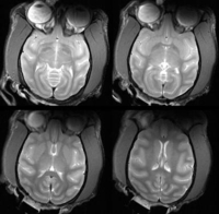
Horizontal sections from a living primate brain. The two eyeballs are visible at the upper edge of the images.
Magnetic resonance imaging (MRI) can be used to visualize the structure of tissues in the brain and other body regions. MRI is a non-invasive method. Images with tissue contrast can be acquired in vivo, i.e. from the living organism.
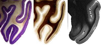
Comparison of two histology images (left and center) with an MRI image (right).
MRI images differ from histological tissue sections in the amount of information that they contain. For one thing, they are three-dimensional rather than two-dimensional. Another advantage is that the contrast can be changed by using different MRI methods. This makes it possible to produce multiple images of the same object with different contrasts to highlight different aspects, for example gray or white matter, the water content of the tissue, or the course of the blood vessels.