


- Electro- physiology
- Measuring how neurons communicate with each other

- Histology
- Tissue sections reveal the cellular structure of the brain
")
- MRI
- Anatomical imaging using tissue contrast
")
- fMRI
- Watching the brain as while works
")
- MRS
- Measuring specific chemicals in the brain

- Surgical procedures
- Operations are always carried out under general anesthesia

- Implants
- Only biocompatible implants made of titanium or special plastics are used

- Alternatives
- Potential and limits of alternatives to animal experiments

Implant Technology
Although today’s implants have excellent biocompatibility and are accepted by the tissue, our laboratory continues to work on improving both the implants and operation techniques.
For example, we fit the implant as specifically as possible to the individual skull shape of the animal in order to speed up the healing process. To do this we actually construct an exact three-dimensional model of the skull surface using MRI images in a sophisticated computer-based process. The implant is then fitted precisely to this model and produced using CNC-assisted milling techniques. The surfaces of the titanium implant are then coated with hydroxylapatite, a component of natural bone substance. In this way we can produce implants that are not rejected by the body, but completely incorporated in the bone structure of the skull.

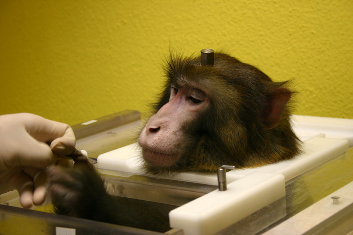
- Rhesus monkey with titanium implant in his chair. Credits: Max Planck Institute for Biological Cybernetics.
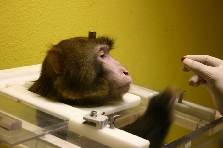

- Rhesus monkey with titanium implant in his chair. Credits: Max Planck Institute for Biological Cybernetics.
No metal parts can be used in animal experiments in the MRI scanner, because they would disturb the magnetic field. In order to avoid such problems we use non-metallic implants out of a special biocompatible plastic for our experiments. This polytheretherketone (PEEK)-based plastic is also used in human medicine. The healing of PEEK implants is as good as that of titanium implants, but until recently there was no method available for coating the plastic with hydroxylapatite. In spring of 2008, however, we made a breakthrough in cooperation with a biomedical engineering company. Just a few months later the first hydroxylapatite-coated PEEK implant was inserted in a procedure filmed by a camera team from the ZDF television channel. The operation scar healed very well; in fact, healing was as good as with titanium implants.
Together with improved (intracutaneous) suture techniques, these innovations in implant materials have substantially shortened the time the animals must spend in a special post-operation chair from around 10 days to only 1 ½ to 3 days.
Together with improved (intracutaneous) suture techniques, these innovations in implant materials have substantially shortened the time the animals must spend in a special post-operation chair from around 10 days to only 1 ½ to 3 days.