


- Electro- physiology
- Measuring how neurons communicate with each other

- Histology
- Tissue sections reveal the cellular structure of the brain
")
- MRI
- Anatomical imaging using tissue contrast
")
- fMRI
- Watching the brain as while works
")
- MRS
- Measuring specific chemicals in the brain

- Surgical procedures
- Operations are always carried out under general anesthesia

- Implants
- Only biocompatible implants made of titanium or special plastics are used

- Alternatives
- Potential and limits of alternatives to animal experiments

Histology
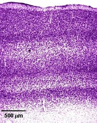
- Tissue section of a macaque brain. This was taken from the brain region responsible for vision. The staining clearly reveals a six-layered structure that is typical of the mammalian brain cortex.

The goal of neuroanatomy is to decipher the organization of the brain.
Histology – the science of biological tissues – plays a major role. With the aid of precision instruments, thin sections of brain tissue are prepared which are only a few thousandths of a millimeter thick. A variety of staining techniques can then be used to visualize specific cells or cell structures. The stained sections are then viewed under the microscope, analyzed and documented.
Histology – the science of biological tissues – plays a major role. With the aid of precision instruments, thin sections of brain tissue are prepared which are only a few thousandths of a millimeter thick. A variety of staining techniques can then be used to visualize specific cells or cell structures. The stained sections are then viewed under the microscope, analyzed and documented.