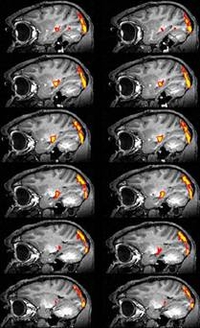
Functional magnetic resonance imaging (fMRI)
Functional magnetic resonance imaging (fMRI) can show the brain at work. Every time we move our little finger or look at a flower, certain regions of the brain are active. And these regions need energy, which reaches the neurons through the blood vessels in the form of oxygen and sugar and is then consumed by the cells. fMRI utilizes this mechanism by visualizing the varying oxygen content of the red blood cells using the so-called “BOLD” (blood oxygen level dependent) effect. A high oxygen content is an indirect indication that the brain cells are active at that location. This method converts the “firing” of the neurons to statistical pictures showing the activation level on a color scale, with yellow symbolizing strong activation and red symbolizing weak activation. If we superimpose the color map on the anatomical MRI image, it is possible to localize neuron activity in a specific anatomical region.
Video: The BOLD effect in Functional MRI
Without stimulation, neurons are generally inactive. As soon as a stimulus in the form of a checker-board pattern (right side) is presented, however, they begin to “fire.” The energy needed for this firing is provided through increased blood flow. The oxygen contained in the blood can be measured on the MRI as the BOLD signal, which provides indirect evidence of brain activity (left side). In the video you can hear the neurons firing; the static noise immediately becomes stronger when the stimulus appears. The BOLD signal, on the other hand, seems to have a lag of several seconds. After the stimulation is discontinued, neuron activity drops markedly (and the static noise decreases), while the BOLD effect persists for a while and only slowly tapers off.
